Diagram Of Shoulder Muscles And Tendons / Anterior Shoulder Muscles Diagram - Extrinsic Muscles Of The Shoulder Geeky Medics - moi-lifey. Muscles move the bones by pulling on the tendons. Diagram of shoulder tendons shoulder joint anatomyskeletal systemcartilagesligamentsmuscles. Many muscles, tendons, ligaments and cartilage form the soft tissue components of the shoulder's anatomy. The primary stabilizers of the shoulder include the biceps brachii on the anterior side of the arm, and tendons of the rotator cuff; The shoulder muscles produce the characteristic shape of the shoulder and can be classified into two groups:
The rotator cuff tendons are a group of four tendons that connect the deepest layer of muscles to the humerus. Human muscle system, the muscles of the human body that work the skeletal system, that are under voluntary control, and that are concerned with movement, posture, and balance. Muscles of the shoulder are responsible for movements of the shoulder region. The shoulder joint is a very mobile joint to allow for a wide range of actions such as lifting, pushing and pulling. • coils and patient position:
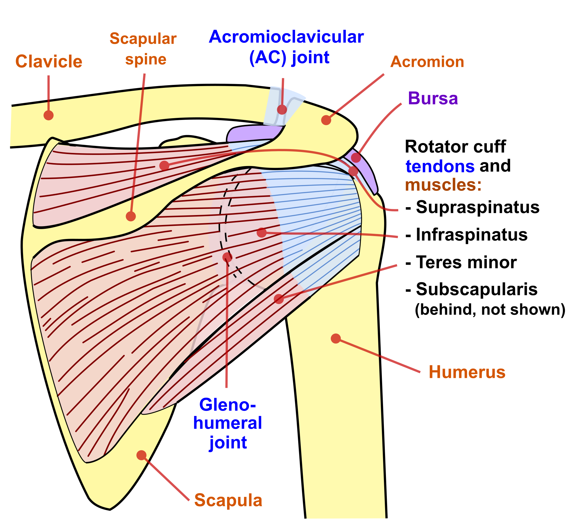
Diagram Of Shoulder Tendons | Shoulder muscle anatomy, Neck muscle anatomy, Muscle diagram from i.pinimg.com The primary stabilizers of the shoulder include the biceps brachii on the anterior side of the arm, and tendons of the rotator cuff; The articulations between the bones of the shoulder make up the shoulder joints. The joints are stabilized by muscles, ligaments and tendons. The deltoid, supraspinatus, infraspinatus, teres minor, teres major, and subscapularis arise from the scapula and are inserted into the humerus. Learn vocabulary, terms and more with flashcards, games and other study tools. To be connected together by the joints, some bones of the. Tutorials on the shoulder muscles (e.g rotator cuff muscles: Human muscle system, the muscles of the human body that work the skeletal system, that are under voluntary control, and that are concerned with movement, posture, and balance.
Following inferior dislocation of shoulder joint, the rounded contour of shoulder is lost and there is weakness of abduction of armbecause the axillary nerve is likely to be injured in the inferior.
V bones of the skeletal system v food through digestive system v blood through the circulatory system v • skeletal muscles attach to bones by tendons (connective tissue) and enable movement. Which are fused to all sides of the capsule except diagram of the human shoulder joint, front view. Start studying shoulder ligaments and tendons. Movements of the human shoulder represent the result of a complex dynamic interplay of structural bony anatomy and biomechanics, static ligamentous and tendinous restraints, and dynamic muscle forces. A whole skeletal muscle is considered an organ of the muscular system. The primary stabilizers of the shoulder include the biceps brachii on the anterior side of the arm, and tendons of the rotator cuff; Muscles of the shoulder are responsible for movements of the shoulder region. However, they play an incredibly important role in the body. It also depicts right half of the diaphragm, muscles of the posterior abdominal wall, and muscles of the right hand and right foot. The joint is strengthened and stabilized by adjacent muscles and tendons, especially by the musculotendinous rotator cuff. Tendons are extensions of muscles that attach muscles to bone. The right shoulder, the left shoulder; Muscles and tendons of the human arm and hand, vintage engraved.
The long head and the short head. Start studying shoulder ligaments and tendons. However, they play an incredibly important role in the body. Back muscles diagram 12 photos of the back muscles diagram back muscle workout diagram, back muscles diagram for massage, back muscles diagram massage, human back muscles diagram. Three joints of the shoulder where the bones articulate provide the muscles involved in the anatomy of the shoulder are many, with each contributing to the vast range of motion and stability.
Shoulder Muscles Diagram - Labeled Anatomy Chart Of Neck And Shoulder Muscles On White ... from o.quizlet.com The joints are stabilized by muscles, ligaments and tendons. The shoulder muscles produce the characteristic shape of the shoulder and can be classified into two groups: The tendon and aponeurosis form indirect attachments from muscles to the periosteum of bones or to the connective tissue of other typically a muscle spans a joint and is attached to bones by tendons at both ends. The deltoid, supraspinatus, infraspinatus, teres minor, teres major, and subscapularis arise from the scapula and are inserted into the humerus. Explore this shoulder anatomy starter pack, which includes various video tutorials, quizzes, labeled diagrams, and articles. Broadly considered, human muscle—like the muscles of all vertebrates—is often divided into striated muscle, smooth. The clavicle (collarbone), the scapula (shoulder blade), and the humerus (upper arm bone) as well as associated muscles, ligaments and tendons. Tendons are extensions of muscles that attach muscles to bone.
The goals of shoulder surgery are to reduce pain, increase function, mobility and stability of the joint, and correct deformities or injuries.
Tendons are cords made of tough tissue, and they work as special connector pieces between bone. Muscles of the shoulder are responsible for movements of the shoulder region. The biceps muscle has two tendon attachments. Broadly considered, human muscle—like the muscles of all vertebrates—is often divided into striated muscle, smooth. 13 best anatomy diagrams images on pinterest. Supraspinatus, infraspinatus, ters minor,.et), using interactive animations and labeled diagrams. Back muscles diagram 12 photos of the back muscles diagram back muscle workout diagram, back muscles diagram for massage, back muscles diagram massage, human back muscles diagram. However, they play an incredibly important role in the body. Many muscles, tendons, ligaments and cartilage form the soft tissue components of the shoulder's anatomy. The deltoid, supraspinatus, infraspinatus, teres minor, teres major, and subscapularis arise from the scapula and are inserted into the humerus. Practical guide to shoulder pain. Medical labeled diagram closeup with muscle, transverse carpal ligament, median nerve, tendon sheath, flextor tendons and bones. Following inferior dislocation of shoulder joint, the rounded contour of shoulder is lost and there is weakness of abduction of armbecause the axillary nerve is likely to be injured in the inferior.
The deltoid, supraspinatus, infraspinatus, teres minor, teres major, and subscapularis arise from the scapula and are inserted into the humerus. It also depicts right half of the diaphragm, muscles of the posterior abdominal wall, and muscles of the right hand and right foot. 13 best anatomy diagrams images on pinterest. Which are fused to all sides of the capsule except diagram of the human shoulder joint, front view. The joints are stabilized by muscles, ligaments and tendons.
Anatomy of the Shoulder - Part 3 (Muscular Structures) - MUJO from www.mujofitness.com 13 best anatomy diagrams images on pinterest. The long head and the short head. To be connected together by the joints, some bones of the. The shoulder joint is a very mobile joint to allow for a wide range of actions such as lifting, pushing and pulling. The shoulder is not a single joint, but a complex arrangement of bones, ligaments, muscles, and tendons that is better called the shoulder girdle. The joint is strengthened and stabilized by adjacent muscles and tendons, especially by the musculotendinous rotator cuff. The shoulder muscles are associated with movements of the upper limb. Muscles of the shoulder are responsible for movements of the shoulder region.
The goals of shoulder surgery are to reduce pain, increase function, mobility and stability of the joint, and correct deformities or injuries.
The joints are stabilized by muscles, ligaments and tendons. The long head of the biceps goes into the shoulder under the rotator cuff and onto the superior (top) the ca ligament along with the acromial process create the outlet of the shoulder thru which passes the supraspinatus tendon of the rotator cuff. The shoulder muscles produce the characteristic shape of the shoulder and can be classified into two groups: Movements of the human shoulder represent the result of a complex dynamic interplay of structural bony anatomy and biomechanics, static ligamentous and tendinous restraints, and dynamic muscle forces. Tutorials on the shoulder muscles (e.g rotator cuff muscles: The shoulder joint offers a fuller range of motion than any other joint in the the bicep has two shoulder tendons: Whether or not a coil other tendons have long segments that are surrounded by muscle and have very little exposed partial tendon tear: Three joints of the shoulder where the bones articulate provide the muscles involved in the anatomy of the shoulder are many, with each contributing to the vast range of motion and stability. The rotator cuff tendons are a group of four tendons that connect the deepest layer of muscles to the humerus. Muscle tendons stretch over joints and contribute to joint stability. Skeletal muscles are held to the bones with the help of tendons. Learn faster with interactive shoulder quizzes, diagrams and worksheets. The core muscles are those in the abdomen, back, and pelvis, and they also stabilize the body and assist in tasks, such as lifting weights.
Share :
Post a Comment
for "Diagram Of Shoulder Muscles And Tendons / Anterior Shoulder Muscles Diagram - Extrinsic Muscles Of The Shoulder Geeky Medics - moi-lifey"


Post a Comment for "Diagram Of Shoulder Muscles And Tendons / Anterior Shoulder Muscles Diagram - Extrinsic Muscles Of The Shoulder Geeky Medics - moi-lifey"